As one of six pulmonologists at Froedtert Pleasant Prairie Hospital, lungs are my specialty. In my work, I see a lot of patients with, for example, COPD (short for chronic obstructive pulmonary disease), asthma, chronic cough, and other diseases and ailments that affect how people breathe. I also have a special interest and training in working with patients who need intensive care.
Many of the patients I see have small, abnormal clumps of cells in one or both lungs that can harden over time to form what we call a lung nodule. Because lung nodules are so small, many patients don't even know they have them. They don't feel them, and they don't have any symptoms. Actually, lung nodules are often discovered accidentally when an x-ray or CT scan of a patient’s arm or shoulder includes part of their chest and reveals something abnormal in one or both lungs.
The vast majority of lung nodules are benign, but occasionally – especially if they continue to grow rapidly over time – they can be an early indicator of lung cancer. When we suspect lung cancer, we need to take a closer look at a patient’s lung nodules to determine what’s causing them and if they are cancerous.
In the past, many of these patients would have had to go to Milwaukee where a cardiothoracic surgeon would perform surgery to examine their lung nodules and collect tissue samples for biopsy, or visit an interventional pulmonologist for special bronchoscopies. But now we perform a much less invasive procedure, right here, close to home, which is much more convenient for patients. The procedure is called a robotic assisted bronchoscopy in which we use a catheter to take biopsy samples from nodules much farther into a patient’s lungs than possible in the past.
With many of these patients, we also want to examine the lymph nodes in the middle of their chest, because that’s often the first place where lung cancer spreads. To accomplish this, we use another minimally-invasive procedure called EBUS – short for endobronchial ultrasound. The EBUS scope goes down into the patient’s trachea to the point just before it divides into both lungs. Then we use the ultrasound on the end of the scope to look on the other side of the airway where the lymph nodes are located. We can also collect a tissue sample for biopsy at the same time.
Often, when we do these procedures, we have the pathologist beside us in the operating room. As we collect tissue for biopsy, we give it directly to the pathologist who can tell us right away whether they see cancer cells or, if not, any indications of infection or other possible causes. In the past, getting that information might have taken a week, but now we’re able to diagnose the ailment and begin treatment much sooner.
Patients still need anesthesia for an EBUS or navigational bronchoscopy, but these procedures are much less invasive. We're just putting thin tubes into the patient’s windpipe. No cutting is happening, so there’s no recovery time. Typically, these patients go home the same day, and are able to resume their normal activities right away.

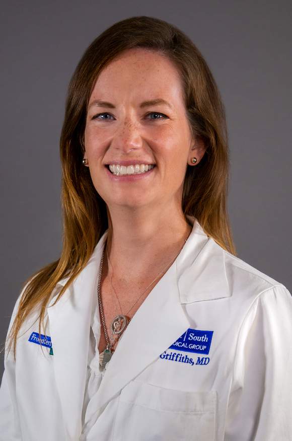
Tricia L. Griffiths, M.D. Pulmonologist & Intensivist, Froedtert Pleasant Prairie Hospital
Many of the patients who come to us with pulmonary nodules and pulmonary masses are long time smokers, have COPD, are on oxygen, and aren’t in the best health. These patients aren’t good candidates for traditional surgery. Putting a frail patient who is in their eighties or nineties through a big surgery to get biopsies from their lungs or lymph nodes might mean a rough recovery for them. But these patients tolerate the less invasive EBUS and navigational bronchoscopy procedures we perform very well. That means we can diagnose them without surgery.
By the time a patient with undiagnosed lung cancer starts having symptoms, their long-term prognosis isn’t very good. But if they get these lung cancer screenings yearly before they have symptoms, we can usually catch things very early and treat them right away.
I recently had a patient who needed both of the less invasive procedures we perform. He was a longtime smoker and had been getting annual CT scans so we could monitor a slow-growing nodule in one of his lungs. When the scan revealed changes in the lung nodule, we performed a navigational bronchoscopy to biopsy it. The results were positive for lung cancer, so we performed an EBUS procedure to see if it had spread to his lymph nodes. Luckily, it had not spread, which means he will likely undergo surgery to remove the nodule. That’s good news for the patient, because it will make it possible for him to go back to work, which is very important to him.
I’d like to see more patients who have smoked in the past take a more active role in monitoring their lung health. Here’s how they can do that: We think of smoking in terms of what we call pack years – defined as smoking one pack of cigarettes per day for one year. For example: if you smoked a half-pack a day for ten years, that’s five pack years. If you smoked a full pack a day for ten years, that’s ten pack years, and so on.
Under the current recommendations, someone who smoked for twenty pack years or more within the last fifteen years, and is between the ages of fifty and eighty, should be getting regular lung cancer screenings. That involves getting a CT scan every year to look for changes in their lungs – including lung nodules or masses.
If you’ve been a longtime smoker, but have never had a CT scan of your lungs, ask your primary care physician - or any of your other doctors - if lung cancer screening is right for you. It’s potentially how you could get yourself on track to keep tabs on your lung health and ensure that you stick around for as long as possible.
Important things to know about lung nodules
According to the American Thoracic Society:
- Lung nodules are found in up to half of adults who get a chest x-ray or CT scan
- Most lung nodules are less than a half-inch in size
- Generally, small lung nodules don’t cause pain or breathing problems
- Fewer than five-percent of lung nodules are cancerous
- If you’re still smoking, quitting is the most important thing you can do to improve your health


